-
Anterior Foot
-
Medial Foot
-
Lateral Foot
-
Sole of Foot
- Step 1. Hover mouse over foot to highlight problem area(s)
- Step 2. Click on highlighted area to list common conditions
- Step 3. Click on condition links for detailed description
Conditions
Click the links below for more info
close
Adult Acquired Flatfoot Deformity (tibalis posterior tendon dysfunction)
Adult acquired flatfoot deformity (also termed posterior tibial tendon dysfunction) is a complex foot and ankle condition characterised by a progressive collapse of the arch with "rolling in" of the ankle. This occurs in adults due to overuse, inflammation and degeneration of the main supporting tendon of the arch called the posterior tibial tendon. The posterior tibial muscle and tendon extends from behind the leg and along the inside the ankle to insert at the top of the arch. This tendon provides a major support to the bone structure of the arch while standing and walking. Overuse of this tendon typically results in pain and swelling behind the ankle and at the top of the arch. Gradually, as this tendon is continually overused, it becomes worn and then weakens. Small tears in the tendon develop and the ankle and arch begin to roll inwards (collapses) resulting in a painful flatfoot. In long standing cases, the tendon can tear completely. .
This condition commonly occurs in patients who are overweight, in their middle age and is more common in females. In the early stages of this deformity, pain is often localised to the inside of the ankle and through the arch. As the foot becomes progressively more flat, the pain can shift from the arch to the outside of the foot and ankle. In the early stages of this condition, the foot is flexible. In long standing cases, the foot and ankle can become arthritic and stiff.
This condition is best treated early, when it is flexible and the arch is yet to completely collapse. If treated early, symptoms can resolve and progression of the flatfoot can be arrested. In most cases, adult acquired flatfoot deformity is generally treated with aggressive conservative care including custom orthotics or ankle bracing, immobilisation in the form of a short leg cast or swalking boot. Immobilisation of the foot and ankle allow the tendon to heal without weight bearing stress on the tendon itself. A structured physical therapy has been shown to help resolve patients’ symptoms in the early stages. Medication such as nonsteroidal anti-inflammatories can help with symptomatic relief of pain, and shoe modifications to a more supportive stiff soled shoes that enable arch supports or bracing of the foot are all important.
Surgery for adult acquired flatfoot deformity is generally considered in cases that do not respond to standard and thorough conservative care. Surgery for adult acquired flatfoot deformity depends on many patient and clinical factors, A painful adult flatfoot can present in many variations, from a mild flexible flat foot to a rigid rolled in ankle. Depending on the presentation, joint preserving surgery versus joint fusion may be warranted. In painful flexible flat feet, bone cuts to realign the foot and tendon transfers and lengthenings can be performed. In stiff painful flat feet, fusion of one or more joints in the rearfoot may be required. In many cases, a mixture of joint fusions and bone cuts to realign the foot are performed together.
close
Ankle Arthritis
Arthritis of the ankle joint can take many forms. It can be localised to a small region of the joint or encompass the entire joint. Localised regions of cartilage damage generally follow an episode of trauma, such as an ankle sprain (see osteochondral lesion of the talus). Another form of joint degeneration can include the production of bone spurs at the front of the ankle (anterior ankle impingement syndrome). These spurs can cause joint impingement and early cartilage wear. These spurs can be painless or patients can experience stiffness, swelling and pain at the front of the ankle joint.
Ankle arthritis can be more extensive and encompass the entire joint. This generally renders the joint painful, stiff and swollen. People who have suffered significant ankle fractures, have rheumatoid arthritis or other inflammatory arthritis can also develop extensive arthritis within the ankle joint.
Treatment for arthritis in the ankle is dependent on many factors, including a patient’s age, their functional capacity, and extent of arthritis in the joint. Treatment such as ice and anti-inflammatory medications can help with symptomatic relief. Other measures such as ankle braces, orthotics and supportive footwear can also help manage symptoms.
Surgical management of ankle arthritis is dependent on the extent and presentation of the underlying arthritis. Isolated bone spurs at the front of the ankle and focal small cartilage lesions can generally be treated with ankle arthroscopy (keyhole surgery). Surgery for more extensive painful arthritis of the ankle joint can range from fusion of the ankle joint, distraction of the joint with an external fixation frame or replacement of the ankle joint.
close
Chronic Ankle Instability
Chronic ankle instability is generally characterised by either mechanical ankle instability or functional ankle instability, or both. Mechanical instability refers to structural damage to the ankle ligaments and joint capsule, rendering the joint unstable. Functional ankle instability refers to the subjective feeling of poor balance and the ankle joint "giving way", particularly on uneven ground.
Chronic ankle instability frequently develops after an ankle sprain (or repeated ankle sprains), where stretching or tearing of the ligaments of the outside of the ankle occur. The ligaments heal in a longer position, so that the ankle ligaments and the ankle joint capsule become slack. The most common signs and symptoms associated with an unstable ankle include repeated ankle sprains, bruising and swelling along the outside of the ankle and foot, persistent pain around the outside and front of the ankle, tenderness with touching the ankle and the feeling that the ankle is unstable.
Treatment of chronic ankle instability is generally achieved with aggressive physiotherapy to improve balance and strength of the muscles around the outside of the ankle. Bracing of the ankle, particularly when playing sport, is very important to help prevent another ankle sprain. Non-steroidal anti-inflammatory medications and icing can help with symptomatic relief of swelling and pain of the ankle.
Should pain and instability of the ankle persist, surgical management may be considered. Treatment of chronic ankle instability includes tightening of the damaged ankle ligaments on the outside of the ankle. In some cases, additional surgery may be required, including debridement of joint synovitis (overgrowth of inflamed scar tissue in the joint capsule), and repair of torn and degenerate tendons on the outside of the ankle (called the peroneal tendons). These additional pathologies commonly accompany chronic ankle instability and multiple ankle sprains.
close
Brachymetatarsia
Brachymetatarsia is a condition in which one of the metatarsal bones in the front of the foot is significantly shorter than the rest. Often the respective toe is shorter and sits in a high position, as if it is floating above the other digits. The most commonly affected bone is the fourth metatarsal. Brachymetatarsia occurs when the metatarsal growth plate closes prematurely and results in a shorter and underdeveloped metatarsal bone.
Brachymetatarsia is generally not painful. Commonly, patients are often concerned with the appearance of their shorter toe. However, people who are active can experience pain in the front of the foot due to the altered weight-bearing pressures across the ball of the foot (due to the short metatarsal).
Surgical intervention for painless brachymetatarsia is not necessary. In cases requiring surgery, lengthening of the short metatarsal is performed.
close
Ganglionic Cyst
Lorem ipsum.
close
Hammertoes
A hammer toe or claw toe deformity refers to buckling and contracture of one or two joints of the lesser toes of the feet. Digital deformity is progressive and begins as mild and flexible and can progress to severe and rigid over time. These deformities can cause pressure from footwear or the ground on the tips of the toes resulting in callus and corn formation. There are many reasons a hammer toe or claw toe deformity can develop. They are commonly associated with bunions due to muscle and ligament imbalances in the forefoot.
Conservative care of lesser digital deformity includes regular podiatry care to debride corns and calluses. In addition, changes to footwear and the implementation of padding or orthotic devices can help. Strapping or splinting of the digits may also be necessary.
Surgical management of hammer or claw toes can be simple or complex. It depends on the overall structure of the forefoot and the flexibility or rigidity of the toe deformity. Straightening of a lesser toe deformity can be performed via minimal invasive surgery. In minimal invasive surgery, specialised instrumentation and techniques are employed to perform tendon, ligament and/ or bone correction through tiny incisions or “keyholes”. In people who have painful corns, boney bumps or crooked toes, minimally invasive foot surgery can work well to resolve these problems without the need for more invasive open surgery. In most cases, minimally invasive surgery can be performed under local anaesthetic without the need for a general anaesthetic and hospitalisation. Mr Smith uses minimally invasive surgery techniques to address common toe and forefoot problems.
Mr Smith also performs more invasive lesser toe surgery, such as fusion of one of the toe joints or via removing a portion of bone from the toe, enabling a toe to be straightened in more complex or rigid toe deformity.
close
Morton's Neuroma
A neuroma is a painful and thickened nerve. This occurs when scar tissue (fibrosis) is formed around a nerve. This process can occur anywhere in the body, including the foot. The most common neuroma of the foot occurs in the webspace between the third and fourth toes (termed Morton's neuroma). Neuromas can form elsewhere, for instance, in the webspace between the second and third toes. The thickened nerve is considered to be a result of compression and continual irritation to the nerve.
Symptoms associated with neuromas include tingling, burning, numbness or sharp-shooting pain into the toes. Additionally, some people feel as if there is something in the ball of their foot, as if they are "walking on a pebble". Commonly, a swelling of a bursa, termed bursitis, occurs overlying a thickened nerve in the webspace. It is not uncommon that this bursitis is more symptomatic than the actual thickened nerve.
Treatment for a Morton’s Neuroma (and accompanying bursitis) includes wearing capacious shoes with a square toe box. This relieves the compression on the forefoot and pressure on the thickened nerve and bursa. Padding or orthotic devices can redistribute pressure from the affected webspace. Activity modifications to avoid repetitive pressure on the forefoot and medications such as non steroidal anti-inflammatories can help with the symptomatic relief of pain.
Injection therapy can result in significant symptomatic improvement, when combined with footwear and activity changes. Injection therapy includes a range of medicines from plain local anaesthetic to corticosteroid or sclerosing alcohol injections. Single or multiple local anaesthetic injections can afford significant early relief of the symptoms of bursitis through laceration of the bursa and stretching of the tissue around the neuroma.
Surgical management of an intermetatarsal neuroma involves removal of the neuroma (and affected nerve) from the webspace of the foot. Alternatively, the nerve can be released, whereby the ligamentous tissue overlying the nerve is cut, similar to decompression of carpal tunnel syndrome in the wrist.
close
Plantar Plate Injury
The plantar plate (syn. flexor plate) is a specialised ligament that is composed of fibrous tissue and cartilage. It is the main stabilising ligament of the joints in the ball of the foot (i.e. where the toes meet the foot) and functions like a ‘pad’. Injury to the plantar plate can be caused by tripping or hyper-extending the toes, but occurs more commonly when the ball of the foot is subjected to daily repetitive weight bearing pressure or overload. This causes attrition of this specialised ligament and eventual tearing. Certain foot types and occupational duties can lead to a gradual overload and rupture of the plantar plate(s). Injury or attrition of the plantar plate is a common cause of hammertoe deformity.
Patients often describe a ‘lump’ or bruise-type discomfort under the ball of the foot (usually under the second digit). It typically becomes worse with prolonged weight bearing and is relieved with rest. Mild swelling may be present, both underneath and on top of the foot. Patients may also notice the second digit gradually moving up or out of alignment.
Depending on the severity of injury, treatment can include rest, massage, padding and strapping. Strapping is considered an important part of treatment. A 6-8 week course of toe-strapping can reduce the severity of hammertoe deformity. Strapping will hold the respective toe in an appropriate position and allow the damaged plantar plate to heal (this process is not unlike strapping of the ankle after ankle ligament injury). Furthermore, orthotics may be required to address foot function causes. In rare cases, walking boots may be considered.
Surgical management of plantar plate injury is varied. Direct repair of the plantar plate (sewing the torn ligament ends together) can be performed. However, a torn plantar plate is often associated with hammertoe deformity and other foot structural problems, such as long metatarsals (these are bones in the fore foot), which commonly need to be corrected to prevent recurrent plantar plate injury from occurring.
close
Tailor's Bunion
A tailor’s bunion or bunionette deformity is similar to a bunion of the big toe joint, except that a tailor’s bunion affects the little toe joint. It involves deviation of the little toe toward the lesser toes and enlargement of the outside of the fifth metatarsal head (where the little toe joins the foot). Tailor’s bunions can cause redness, swelling and pain due to footwear irritation over the prominent bump. The term tailor’s bunion was coined following the common presentation of these deformities in tailors who would sit cross-legged and put pressure on the outside edge of their foot. This constant rubbing led to the bump at the base of the fifth toe.
Tailor’s bunions are generally considered to be caused by either a family predisposition or foot function. The diagnosis of the tailor’s bunion is generally apparent upon inspection of the foot – a lump can be seen on the side of the little toe joint.
The treatment of tailor’s bunions generally involves attention to footwear that does not rub over the bump of the fifth toe. Medication, such as non-steroidal anti-inflammatory drugs, can afford symptomatic relief. Padding over the area can help reduce the pressure over the bump.
close
Adult Acquired Flatfoot Deformity
Adult acquired flatfoot deformity (also termed posterior tibial tendon dysfunction) is a complex foot and ankle condition characterised by a progressive collapse of the arch with "rolling in" of the ankle. This occurs in adults due to overuse, inflammation and degeneration of the main supporting tendon of the arch called the posterior tibial tendon. The posterior tibial muscle and tendon extends from behind the leg and along the inside the ankle to insert at the top of the arch. This tendon provides a major support to the bone structure of the arch while standing and walking. Overuse of this tendon typically results in pain and swelling behind the ankle and at the top of the arch. Gradually, as this tendon is continually overused, it becomes worn and then weakens. Small tears in the tendon develop and the ankle and arch begin to roll inwards (collapses) resulting in a painful flatfoot. In long standing cases, the tendon can tear completely.
This condition is best treated early, when it is flexible and the arch is yet to completely collapse. If treated early, symptoms can resolve and progression of the flatfoot can be arrested. In most cases, adult acquired flatfoot deformity is generally treated with aggressive conservative care including custom orthotics or ankle bracing, immobilisation in the form of a short leg cast or walking boot. Immobilisation of the foot and ankle allow the tendon to heal without weight bearing stress on the tendon itself. A structured physical therapy has been shown to help resolve patients’ symptoms in the early stages. Medication such as non-steroidal anti-inflammatories can help with symptomatic relief of pain, and shoe modifications to a more supportive stiff soled shoes that enable arch supports or bracing of the foot are all important.
Surgery for adult acquired flatfoot deformity is generally considered in cases that do not respond to standard and thorough conservative care. Surgery for adult acquired flatfoot deformity depends on many patient and clinical factors, A painful adult flatfoot can present in many variations, from a mild flexible flat foot to a rigid rolled in ankle. Depending on the presentation, joint preserving surgery versus joint fusion may be warranted. In painful flexible flat feet, bone cuts to realign the foot and tendon transfers and lengthening can be performed. In stiff painful flat feet, fusion of one or more joints in the rearfoot may be required. In many cases, a mixture of joint fusions and bone cuts to realign the foot are performed together.
close
Fifth Metatarsal Fractures
The fifth metatarsal is a bone on the outside of the midfoot region. It is one of the most commonly broken bones in the foot. Injury to the foot, including an ankle sprain or falling over a gutter or a stair can result in a fracture to this bone. In addition, training errors can result in a fracture of the fifth metatarsal.
Generally speaking, all fractures of the fifth metatarsal results in bruising, pain, swelling and tenderness along the outside of the foot. They can be painful to walk on and generally require treatment. Initial treatment is aimed at reducing swelling and immobilising the foot to allow bone to heal. This includes ice therapy, compression and immobilisation in a cast or walking boot. Certain fractures are prone to delayed healing require a below knee cast and non-weight bearing on crutches. If the broken bone is significantly displaced or has failed to heal following conservative treatment, surgery may be required. Surgery for broken bones varies depending on the type of fracture, the position of the bone and can include a screw, a plate or a wire.
close
Ganglionic Cyst
A ganglion (ganglionic cyst) is an out-pouching from a tendon sheath or a joint capsule.
Ganglions are very common and frequently occur in the foot. They can often fluctuate in size and resolve spontaneously without treatment. There is debate over the origins of a ganglion, however it is generally believed that a weakness or tear in the lining of a joint capsule or a tendon sheath allows for the escape of fluid into a balloon like mass. Ganglions can become painful if they are large, subjected to pressure from footwear or a bony prominence and if they reside next to a nerve.
Treatment for ganglions varies from watchful waiting to surgical excision. Shoe modifications or padding may help redistribute pressure away from the ganglion. A common clinical treatment involves aspiration (drawing out) of the fluid within the ganglion and injecting steroid into the area to help prevent its recurrence. However, ganglions can recur despite this treatment and often several courses of aspiration may be required. Surgery to remove the ganglion can be considered if conservative options fail.
close
Tarsal Coalition
A tarsal coalition is an abnormal join or connection between two bones in the foot. This commonly occurs in the tarsal bones of the foot (in the rearfoot). This abnormal connection can involve fibrous or cartilage tissue, or it can be a complete bony join. A tarsal coalition generally occurs during foetal development, whereupon the individual bones do not mature fully. The two most common coalitions in the foot are between the calcaneus and the navicular and the calcaneus and the talus. The calcaneus is the heel bone. The talus sits on top of this, just below the ankle. The navicular bone resides at the top of the arch of the foot. Coalitions or pseudo-coalitions can also occur following trauma or infection.
Not all coalitions are painful. However, a non painful coalition can become painful following trauma, such as rolling an ankle. This type of injury can disrupt a non painful coalition, resulting in pain. Generally, coalitions become painful during childhood and adolescence as children begin to become more active and this places stress through a coalition, causing pain. Common symptoms of tarsal coalitions include swelling and pain in the rear foot or ankle that occurs with prolonged walking or activity. Muscle spasms on the outside of the leg with prolonged walking can occur in certain cases. A stiff flat foot is a common presentation in children with a tarsal coalition.
Conservative management of tarsal coalitions is aimed at relief of pain. Oral non-steroidal anti-inflammatory medications can provide temporary relief. Corticosteroid injections into the painful region can help with acute symptomatic relief. Custom orthotic devices can be beneficial in distributing weight away from the respective coalition. Periodically, below knee cast immobilisation is required to avoid placing weight on a painful foot.
Surgical management of tarsal coalition is dependent on presentation. In juvenile and adolescent cases, removing the coalition generally provides for long term symptomatic relief. In the adult population a tarsal coalition can generally result in joint arthrosis and fusion (arthrodesis) of the affected joint in the rear foot is generally required.
close
Talar Dome Lesion
A talar dome lesion (termed osteochondral lesion of the talus) refers to a focal area of cartilage degeneration on the talus (the bone that makes up the inferior portion of the ankle joint). The top of the talus is dome shaped and covered in thick cartilage. Talar dome lesions are generally caused by injury, such as an ankle fracture or severe ankle sprain. This damage results in a small crack in the cartilage, which can degenerate into a loosening of the cartilage and escape of joint fluid underneath this. This fluid can penetrate the bone and form a bone cyst. Sometimes the cartilage can become soft, break off and float within the joint.
These injuries are very subtle and may not produce symptoms for many months or years after the initial injury. Common signs and symptoms of an osteochondral lesion of the talus include pain deep in the ankle, especially with weight bearing and going up and down stairs. In some patients their ankle can click and "catch" or "lock up" when they walk. Episodes of swelling in the ankle is common.
Conservative treatment of talar dome injuries includes oral anti-inflammatory medications and icing for symptomatic relief. Physical therapy involving range of motion exercises and strengthening of the peroneal tendons (ankle stabilisers) can help when a talar dome lesion is associated with ankle instability. Immobilisation in the form of walker boot may help symptoms.
Surgical management is often required if conservative care fails to relieve symptoms. Removal of any loose fragments is necessary, followed by drilling or "micro fracture" of the cartilage lesion "bed" to promote cartilage healing. Cartilage healing occurs with a secondary type of cartilage, termed fibrocartilage, which fills the lesion of the talar dome. This can generally be done via an arthroscope or open procedure.
close
Bunions (Hallux Valgus)
The term bunion (or hallux valgus) is used to describe a structural deformity of the big toe joint, specifically deviation of the big toe towards the lesser toes and deviation of the first metatarsal towards midline of the body, leading to subluxation of the big toe joint. A common misconception is that a bunion is a bony growth on the inside of the big toe joint. This is not true, as the bump on the side of the big toe joint is the joint itself protruding outwards (as it becomes subluxed). A bunion is a progressive deformity and may lead to arthritis of the big toe joint, especially in people over 50 years of age. It is the most common condition affecting the big toe and its prevalence increases with advancing age affecting up to 65% of the population over 65 years of age. Bunions have been found to have a negative impact on a person's health related quality of life.
It is generally accepted that bunions are inherited (i.e. a predisposition to develop them) and recent research supports this. Footwear may hasten the development of a bunion, but is generally not considered the primary cause. Bunions can affect children and this is termed juvenile hallux valgus. This is usually due to the position of cartilage in the big toe joint.
Bunions can become painful for several reasons. The bump can rub on footwear and become inflamed and painful. In addition, the mal-aligned joint can led to arthritis. People with bunions can experience a deep ache and throbbing sensation within the joint as a result of this arthritis.
Surgery has been found to be superior for patient outcomes, including quality of life and pain, when compared to orthotics (arch supports) and watchful waiting. However, symptomatic improvement can be achieved in certain cases where footwear changes, padding around the bump and activity modifications decrease stress through the big toe joint and irritation over the bump. Anti-inflammatory medications can help with symptomatic pain and orthotic devices can provide some early short-term relief, particularly in feet that are flat.
There are over 150 different types of procedures reported to fix bunions. Generally speaking, in the hands of an experienced foot surgeon and depending on the severity of the bunion, only two or three different types of procedures are used. Surgical management involves bone cuts to realign the big toe joint combined with balancing of the soft tissue around the joint. This restores the appropriate alignment of the joint and position of the toe. In certain cases, where there is extensive arthritis in a bunion, for example in rheumatoid arthritis or gout, fusion of the big toe joint is a more predictable and reliable long-term procedure.
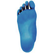
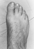
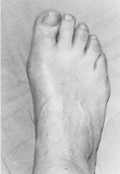
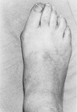
How Big Is Your Bunion?
A common classification scheme for bunions is referred to as the Manchester scale. This scale utilises a standardized set of clinical photographs of feet with 4 stages of bunion severity: (1) None, (2) Mild, (3) Moderate and (4) Severe. This classification scheme has excellent reliability and is a valid tool for grading of severity of bunions and for clinical and research applications.
(1) No Bunion
(2) Mild
(3) Moderate
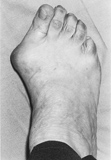
(4) Severe
Reproduced with permission, from: Journal of the American Podiatric Medical Association. From the article: Garrow AP, Papageorgiou A, Silman AJ, Thomas E, Jayson MIV, McFarlane GJ. The grading of hallux valgus: The Manchester scale. Journal of the Podiatric Medical Association, 91:74-78, 2001.
close
Hallux Rigidus
Arthritis of the big toe joint (termed hallux rigidus) is a condition characterised by pain, swelling and stiffness of the big toe joint. Big toe joint arthritis is generally caused by poor foot function or from trauma i.e. badly stubbing a toe. Both result in a cartilage damage within the joint and inflammation of the joint capsule. As the cartilage damage progresses over a period of time, bone spurs form around the joint and it becomes progressively more stiff. Big toe joint arthritis can also be caused by inflammatory arthritis such as rheumatoid arthritis or gout.
This condition can cause difficulty with daily activities, such as walking and running. Like all arthritis it can be aggravated by cold and damp weather. In addition, lumps that develop around the top of the joint can make certain footwear difficult and painful to wear.
Shoe modifications, such as a spacious toe box or stiff rocker-bottom soles may be necessary. Orthotic devices to improve foot function or to limit the motion of the big toe can also help. Medications such as nonsteroidal anti-inflammatories or paracetamol may help with symptomatic relief. Supplements such as glucosamine and fish oil may prove helpful. Injections into the joint of corticosteroids can help afford short term relief, but are generally not recommended.
Surgical management of this condition is dependant on the extent of arthritis in the joint. If there is only a small amount of arthritis in the joint and pain is related to spurring around the top of the joint, then removal of the bone spurring around the joint can provide symptomatic relief. In more advanced cases, where there is more extensive cartilage wear and stiffness in the toe, fusion (termed arthrodesis) of the big toe joint is necessary. Often, patients are reluctant to consider fusion of the big toe joint, however, this procedure essentially creates a painless stiff toe (from a painful stiff joint!).
close
Seasamoid Injuries
The sesamoid bones are small oval shaped bones that reside under the big toe joint. These sesamoid bones are enveloped in tendons and provide attachments for ligaments and the joint capsule around the big toe.
Sesamoid injuries can range from simple inflammation to fracture of the bone. Injuries are more common in people with a high arched foot as they place higher pressure on the ball of their foot. In addition, sesamoid injuries commonly occur in certain sporting activities such as running, basketball, tennis and dancing. High heel shoes can also overload the ball of foot and predispose to sesamoid overload and pain.
A common sesamoid pathology is termed sesamoiditis. This is an overuse injury that results in inflammation of the sesamoid bones and the tendons that attach into these. Signs and symptoms include swelling and dull pain directly beneath the big toe joint. The pain is generally episodic and follows sporting activities or with wearing certain footwear, such as high heels.
A less common sesamoid injury is termed osteonecrosis. This occurs when the sesamoid bone becomes unhealthy and degenerates due to interruption to the blood flow to the sesamoid. This can occur in chronic cases of sesamoiditis and overload to the area. It can also occur following a fracture. Signs and symptoms include a dull pain with symptomatic swelling beneath the big toe joint.
A sesamoid bone can become fractured following acute trauma or repeated overload. This generally produces immediate swelling and bruising of the area and can be extremely tender to walk on.
Treatment for sesamoid injuries needs to be aggressive. Orthotics with forefoot padding to off load the sesamoid area is a necessity for people who have a high arch or involved in sports that place an increased pressure underlying the big toe joint. In the short term, padding and strapping can help to protect the sesamoid region. Oral anti-inflammatory medications and ice can provide symptomatic relief. In more severe cases immobilisation in a walking boot or non weight bearing fibreglass cast in may be warranted. Steroid injections are generally not recommended.
When fractured or degenerate sesamoids remain painful and fail to heal, excision of the fractured or degenerate sesamoid may be necessary.
close
Haglund's Deformity
Haglund’s deformity is a bony lump that grows on the back of the heel bone, which can be irritated by footwear. In some cases a bursa (or sac of fluid) in this area can become inflamed and painful. This condition is common in people with a high arch foot or a tight Achilles tendon. Symptoms can include a pain in the back of the heel, swelling, warmth and redness over the lump.
Treatment for Haglund’s deformity is aimed at relief of pain, resolution of inflammation and redistribution of pressure away from the bump. This includes ice and anti-inflammatory medication which can help with symptomatic relief of pain. Exercises to stretch the Achilles tendon and heel lifts in the shoes can help. Padding in footwear and shoe modifications can also avoid irritation to the bump. In some instances immobilisation in a walking boot may be necessary to settle acute flares in symptoms. Surgical management of Haglund’s deformity is only required if symptoms persist and involves excision of the bony bump at the back of the heel.
close
Heel Pain (Plantar Fasciitis)
Inferior heel pain (termed insertional plantar fasciitis) is one of the most common foot disorders. Generally, inferior heel pain is caused by several degenerate processes to the structures of the inferior heel region, including a stress response of the heel bone, degeneration of the attachment of a ligament into the heel bone (this ligament is termed the plantar fascia) and swelling within the small foot muscles in the sole of the foot. In long standing cases, tightening of the soft tissue around the heel can occur and cause tethering of a nerve in this region.
Statistically, the most common finding in people who have inferior heel pain is a high body mass index (people who are obese or overweight). Secondly, people involved in weight bearing occupations on hard concrete or flat surfaces are also predisposed to this problem.
Common symptoms of inferior heel pain include a bruise or throb-like pain underneath the heel of the foot. This can be particularly pronounced after rest or with getting out of bed in the morning. In addition, the heel can also become progressively sore with prolonged weight bearing throughout the day. Pain is generally localised underneath the heel and around the periphery of the heel. In some cases, pain can extend to the plantar fascia ligament in the arch and to the muscles on the inside of the heel. When a nerve entrapment occurs, this can cause sharp shooting pains into the leg and along the outside of the foot.
Commonly, x-rays demonstrate a heel spur. This heel spur is not considered the focal source of a patient’s pain, although its presence can dictate chronic episodes of bone stress and bone healing. In certain cases, the spur can fracture and become a source of pain.
Treatment for inferior heel pain and plantar fasciitis includes stretching exercises of the calf and foot. Massage of the calf muscles and arch muscles is important. Attention to wearing supportive lace-up footwear with good shock absorption is recommended. Anti-inflammatory medication and ice can provide symptomatic relief. Activity modifications and occupational modifications may be necessary in order to promote resolution. Commonly, arch supports or custom made orthotic devices result in significant improvement and resolution of inferior heel pain. If heel pain persists, a single or multiple injections with a local anaesthetic or corticosteroid can provide symptomatic relief. In most severe cases a removable walking boot or night splint may be worn to help rest the area. In very chronic cases, extracorporeal shock wave therapy into the inferior heel can provide relief.
It is important for each patient to remember that the predisposition to developing inferior heel pain overwhelmingly has been shown to be increased weight. Therefore, before any surgical treatment is considered, weight reduction tactics are paramount.
The need for surgical management of inferior heel pain is not common. Releasing the plantar fascia ligament and tight soft tissue is a common procedure to address inferior heel pain. In addition, recent advances using "radio-frequency coblation", where a probe is inserted repeatedly into the plantar fascia ligament to create an specific type of acute injury (thereby stimulating a healing response) is a means of providing for a more functional postoperative course and outcome.
close
Achilles Tendon Rupture
The Achilles tendon is the strongest tendon in the human body. It courses behind the heel and attaches into the heel bone. Tears of the Achilles tendon are can commonly occur with walking down stairs or stepping off of a gutter to cross the road. Tears can also occur from jumping or changing direction suddenly. Less commonly, it can be caused by falling or tripping.
Tears of the Achilles tendon can be partial or complete. Complete tears commonly occur directly behind the ankle in the mid portion of the tendon. Signs and symptoms associated with rupture of the Achilles tendon include a popping or cracking sound. People experience sudden onset of pain and describe a feeling as if they were kicked in the back of the leg. There is generally immediate bruising and swelling around the ankle and along the outside of the foot. People have difficulty with walking due to the loss of power of the muscles in the back of the leg.
It is very important that an early diagnosis of ruptured Achilles tendon is made. Inappropriate early treatment or delayed attention to ruptured Achilles tendon can result in long term weakness in the affected side. As with many soft tissue injuries, the appropriate early treatment should be rest, ice, compression and elevation. However, there is debate over non-surgical treatment versus surgical treatment for the management of a complete rupture of the Achilles tendon.
Non-surgical treatment includes a below knee cast with the foot in a pointed position to allow the torn tendon ends to get as close to each other as possible to begin healing. This generally involves several months of casting with crutches non weight bearing to weight bearing in a walking boot with heel lifts. Physiotherapy and active range of motion exercises with braces are often also employed to allow for improved healing of the tendon and to gain strength. There is a higher rate of recurrence of Achilles tendon tears when they are treated conservatively. However, there is a lower complication rate compared with surgery.
Surgery for a complete rupture of the Achilles tendon significantly decreases the risk of re-rupture of the Achilles tendon. Surgery includes joining the torn ends of the tendon with strong suture material to provide for stable repair. Following surgery, the foot is generally immobilised for two weeks with the foot pointed down whereupon the patient can then begin walking with protected weight bearing in a walking boot with heel lift. Physiotherapy and range of motion exercises are started early to improve function and strength.
Partial tears of the Achilles tendon generally occur higher up in the leg closer to the muscle where they have good blood flow. These partial ruptures are most often managed conservatively in a walking boot with physiotherapy and generally heal uneventfully.
close
Achilles Tendinopathy
The Achilles tendon is the strongest tendon in the human body. It is the tendon that joins the calf muscle to the heel bone at the back of the ankle. There are several common conditions that can affect the Achilles tendon and cause pain along the back of their ankle and heel. These conditions can involve the tendon itself, the insertion of the tendon into the heel bone or the lining of the tendon. Sometimes both the lining of the tendon and the tendon can become damaged. In some cases, the tendon can completely rupture.
Damage to the lining of the Achilles Tendon (paratenonopathy)
Inflammation of the lining of the tendon (called the paratenon) can occur from overuse of the tendon and generally follows a sudden increase in activity or with the beginning a new exercise program. Other contributing factors include: walking on inclined treadmills or hills, tight calf muscles, limb alignment and foot function problems and certain medication (fluoroquinolone antibiotics). This puts too much stress through the tendon and results in pain and inflammation of the lining of the tendon. Aching, stiffness and tenderness along the tendon behind the ankle is common. In severe cases, the tendon can be swollen and a crackling sound occurs when the foot is moved up and down. This crackling occurs as a result of adhesion of the paratenon to the Achilles tendon. In chronic cases, the Achilles tendon can also become damaged and thickened and this called tendinosis (see below). People can suffer from paratenonopathy and tendinosis simultaneously.
In the early to middle stages, treatment includes immobilisation in the form of a walking boot. Ice therapy and oral medication, such as non-steroidal anti-inflammatories, can help early symptomatic relief and prevent inflammation from becoming chronic. Orthotics can help with elevating the heel and taking tension off the tight and painful Achilles tendon. Night splints can be quite helpful to maintain a mild stretch on the Achilles tendon during sleep so that it does not become stiff and contracted overnight. Stretching of the tendon and physiotherapy is important for the management of this condition. Physical therapy includes massage and strengthening exercises, ultrasound therapy and progression to strengthening exercises of the Achilles tendon. Other treatments under investigation include local anaesthetic or sclerosing injections, shock wave therapy, iontophoresis, topical glyceryl trinitrate and low-level laser therapy.
Damage to the Achilles Tendon (Achilles Tendinosis)
Tendinosis of the Achilles tendon can occur at two regions, (1) in the tendon itself, behind the ankle and, (2) at the insertion of the Achilles tendon into the heel bone. These are two separate entities, but have the common findings of degeneration of the tendon fibres and the development of scar tissue and ingrowth of new blood vessels. This commonly results in a lump within the tendon, which can be easily identified and felt at the back of the ankle. At the insertion of the tendon, the production of new cartilage and calcification results in bone spur formation. Both of these conditions can be very debilitating and generally represent a chronic form of tendon degeneration. Inflammation and scarring of the lining of the tendon (paratenonopathy) can also occur with this condition (see above).
Tendinosis can occur from overuse of the tendon and generally follows a sudden increase in activity or with the beginning a new exercise program. Other contributing factors include, walking on inclined treadmills or hills, tight calf muscles, limb alignment and foot function problems and certain medication (fluoroquinolone antibiotics). This puts too much stress through the tendon and results in degeneration of the tendon. Aching, stiffness and tenderness along the tendon with a palpable lump behind the ankle is common.
Treatment for tendinosis of the Achilles tendon includes all the aforementioned modalities for paratenonopathy. Most importantly, structured sequential treatment of this condition is required. This includes immobilisation and heel lifts, night splints and physiotherapy, followed by a gradual strengthening program. In non responsive cases, autologous blood injections, shock wave therapy or sclerosing agents can prove successful.
Surgical management for tendinosis in the region of the Achilles tendon belly includes excision of the scar tissue or damaged tendon and sewing the tendon back together. For tendinosis at the attachment of the tendon in the heel includes partial detachment of the Achilles tendon insertion and debridement of the scar tissue, calcifications and bone spurs.
close
Peroneal Tendon Injuries
The peroneal tendons comprise two tendons that run along the outside of the ankle. One tendon inserts onto the outside border of the foot (on the fifth metatarsal) and another one travels underneath the foot (and inserts at the base of the 1st metatarsal). These tendons are involved in stabilising the foot during normal walking.
A common cause of a peroneal tendon injury occurs following an ankle sprains. This tendon injury is often overlooked following a sprain and can be the source of continued pain in the lateral ankle. In addition, people who have high arched feet and who tend to “roll out” when they walk place more stress along these tendons and are predisposed to overuse injury.
There are several types of peroneal tendon injuries. Inflammation of the peroneal tendons and their tendon sheath can involve pain and swelling along the course of the tendon. The peroneal tendons can split behind the ankle bone as one tendon tends to compress another against the ankle bone. This causes pain, swelling and weakness of this muscle group. In cases of severe ankle sprains the peroneal tendons can also slip out of their normal groove, (termed peroneal subluxation). When this occurs, a snap or popping sound can be heard as these tendons roll over the ankle bone and sublux out of their groove. This can often result in swelling and pain and adds to ankle instability or weakness.
Conservative treatment of peroneal tendon disorders can include immobilisation, ice and anti inflammatory medications to help with symptoms. Ankle bracing and physiotherapy can help resolve and prevent recurrence of peroneal tendonitis. Orthotics can help to balance high arched foot type.
Surgical management depends on the type of peroneal tendon pathology. Debridement of damaged peroneal tendons and repair of any splits within the tendon can resolve peroneal tendon pain. Subluxation of the peroneal tendons can require tightening of the tendon sheath or remodelling of the groove behind the ankle bone to prevent subluxation.
close
Os Trigonum Syndrome
An Os trigonum is a small accessory bone that sits behind the ankle joint. It is connected to a bone that forms part of the ankle joint, called the talus, by a cartilage or fibrous band. Extreme flexion of the foot downwards causes an impingement of this bone behind the ankle, like a nutcracker. In certain cases, impingment of the tendon to the big toe by the Os trigonum can also occur. Os trigonum syndrome occurs commonly in athletes, particularly soccer players, footballers and dancers.
The signs and symptoms of Os trigonum syndrome include aching and throbbing in the back of the ankle. This most commonly occurs with significant flexion of the ankle or pushing down with the big toe when walking. In some cases, swelling in the back of the ankle is present.
The treatment of Os trigonum syndrome generally involves rest and immobilisation in a walker boot to allow for injured tissue to mend. Ice and anti-inflammatory medications can provide symptomatic relief. In certain cases, corticosteroid injections into the posterior ankle region can help reduce inflammation and pain. Surgical management generally involves excision of the Os trigonum. In addition, debridement or decompression of the tendon to the big toe, which courses behind the ankle joint in close proximity to the Os trigonum, is required.
close
Painful little toes
Painful corns can grow on the inside or outside of the 5th toe. This is a very common foot problem and can be a source of long term frustration for patients. Painful corns most commonly occur in people with crooked or stiffened little toes, such as claw toes or hammertoes. Corns are a focal collection of densely compact skin cells. Pressure from shoes against a stiffened or crooked 5th toe can result in the growth of corns. Regular removal or podiatry care of the painful corn can provide temporary relief, but the corn will often return. Corrective toe surgery may be required in instances where toe deformity contributes to corn formation. Surgical management of the 5th toe can be performed with the aim of permanently removing the corn.
Straightening of a small toe to manage painful corns or ulcers can be performed via minimal invasive surgery. In minimal invasive surgery, specialised instrumentation and techniques are employed to perform tendon, ligament and/ or bone correction through tiny incisions or “keyholes”. Minimally invasive foot surgery can work well to resolve little toe problems without the need for more invasive open surgery. In most cases, minimally invasive surgery can be performed under local anaesthetic without the need for a general anaesthetic and hospitalisation.
Mr Smith uses minimally invasive surgery techniques to address common toe and forefoot problems.
close
Ingrown Toenails
Ingrown toenails (also known as onychocryptosis) are one of the most common foot complaints. Ingrown toenails occur when the nail plate aggravates the surrounding nail fold or skin. They can cause intense pain, redness and swelling, and can lead to infection.
The great toe is the most common site for ingrown toenails; however, they can occur on any of toe. Some of the more common causes for ingrown toenails to develop are: trauma or injury to the nail; the structural shape of the nail; poor cutting technique; digital deformities; and ill-fitting footwear.
Surgical management for ingrown toenails is one of the most common foot surgeries performed in Australia. Mr Simon Smith has dedicated more than 10 years to the clinical supervision and instruction of nail surgery techniques to undergraduate podiatry students at the La Trobe University Health Sciences Clinic.
Surgical management of ingrown toenails is aimed at providing permanent resolution of the ingrown toenail. The most common procedures performed for the management of ingrown toenails includes removing a small portion of nail (partial nail avulsion) and cauterisation of the nail root, or via sharp excision of the nail and nail root with stitches.